Biology is a longstanding frontier for quantum metrology, where high optical intensities are frequently required to get sufficient signal, but lead to damage in the biological sample. Quantum optics based precision measurements allow extracting the most information from the light emitted by the sample without using high intensities. Our lab aims to translate the precision sensing techniques from quantum optics to biological measurements. The efforts in this lab can be separated into categories: 1) quantum microscopy, 2) quantum limited biosensing, 3) deep tissue imaging, 4) optical tweezers, and 5) life microscopy.
Biosensing
In biology at microscale, weak optical signals need to be detected in large level of background noise. A straightforward solution to this issue is to increase the probing optical intensity interrogating the sample. However, high optical intensities are known to induce photodamage to biological samples, which can affect their viability, growth and function. This greatly limits the sensitivity of biosensors and microscopes, preventing the observation of rapid biological phenomena or processes that happened at smaller scales. Therefore, we aim to translate the precision sensing techniques from quantum optics to biological measurements. This demonstrates improved sensitivity and measurement speed of biosensors, which provide a path towards the observation of new biophysical phenomena at safe intensities for extended periods of time.
Recent Work
Quantum Biotechnology
This is the first comprehensive review in quantum biotechnology to integrate the traditionally separate disciplines of quantum physics and biotechnology, with a focus on four emerging frontiers: quantum-enabled sensing, quantum-enabled imaging, quantum biomolecular control, and quantum effects in biological systems.
Read more:
N. Mauranyapin, et al., Quantum Biotechnology, Advanced Quantum Technologies, pp. 2100139 (2022).
Interferometric Biosensing
This book chapter provides an overview of interferometry-based biosensing techniques, covering recent advances, core principles, experimental methodologies, and their applications in protein detection and dynamic analysis at the single-molecule level.
Read more:
N. Mauranyapin, et al., Interferometric Biosensing, Single Molecule Sensing Beyond Fluorescence, Springer International Publishing, pp. 5-36 (2022).
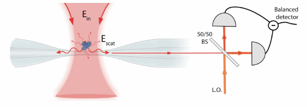
Biosensing at the quantum noise limit
Biosensors that are able to detect and track single biomolecules without requiring labelling are an important tool to understand biophysics including biomolecular dynamics and interactions. They are valuable tools for medical diagnostics and need to be operated at their ultimate detection limits. We have developed a biosensor free of all technical noise sources that operates at the fundamental precision limit due to the quantization of light. This demonstrated state-of-the-art sensitivity using only a tiny fraction of power compared to other sensors (four orders of magnitude reduction in optical intensity), and thereby greatly reduces photodamage dealt by detection. Our approach provides a pathway towards quantum-enhanced single-molecule biosensors by achieving quantum noise-limited precision.
Read more:
N. Mauranyapin, et al., Evanescent single-molecule biosensing with quantum-limited precision, Nature Photonics, 11, 477–481 (2017).
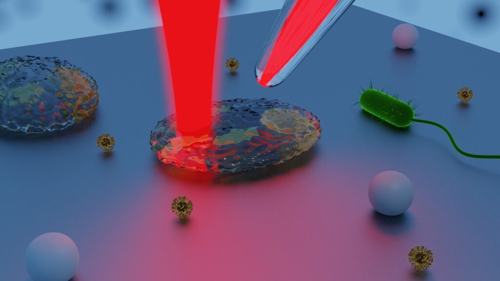
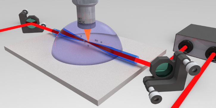
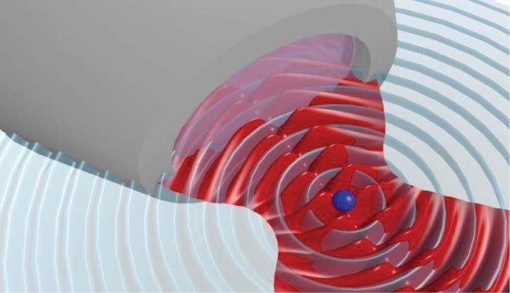
Quantum enhanced optical tweezers
Optical tweezers are a common tool in biological experiments that use high intensity, tightly focused beams to manipulate and track single cells and small glass spheres in live biological samples. For the first time, we have demonstrated that the fundamental limit due to the quantization of light can be overcome for measurements of living systems. This laser-based microparticle tracking technique is compatible with non-classical light and is immune to low-frequency noise, leading the way to achieving a broad range of quantum-enhanced measurements in biology.
Recent Work
Ultrafast viscosity measurement with ballistic optical tweezers
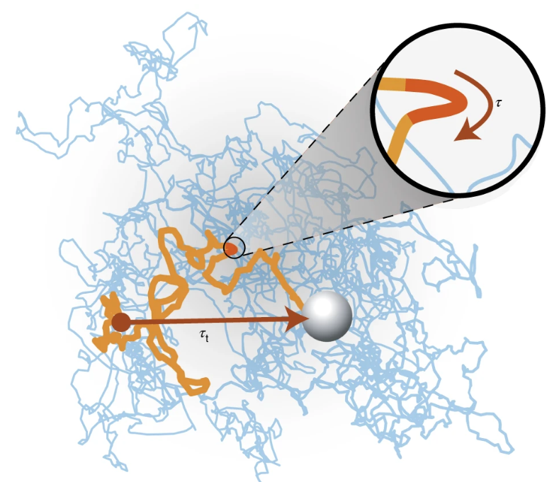
We have developed a new technique for ultrafast viscosity measurement using optical tweezers, enabling real-time tracking of fluid viscosity with temporal resolution as short as 20 μs. By resolving the instantaneous velocity of a trapped particle using a structured-light detection scheme, we achieved measurement speeds over four orders of magnitude faster than existing methods, while using standard optical components. This approach allows viscosity to be treated as a dynamic property, sensitive to rapid changes in the local environment. We hope it will offer a useful tool for exploring fast dynamics in biological and soft-matter systems.
Read more:
L. Madsen, et. al., Ultrafast viscosity measurement with ballistic optical tweezers, Nature Photonics, 15 386-392 (2021).
See also: M. Taylor, et. al., Biological measurement beyond the quantum limit, Nature Photonics, 7 229-233 (2013).
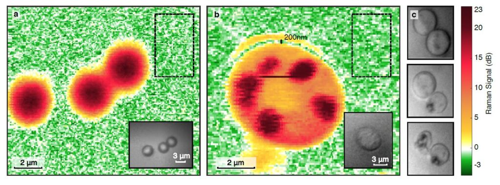
Quantum Enhanced Raman Microscopy
Raman microscopy allows measurements on biological samples where not only the contrast of the sample, but also the chemical composition is visible. However, photodamages introduce hard limits on performances that theory predicts which can only be overcome using quantum photon correlations. We experimentally validate this using quantum correlated light to reach signal-to-noise beyond the photodamage-free capacity of conventional microscopy. Our work provides a path towards order-of-magnitude improvements in both sensitivity and imaging speed.

Recent Work
Fast biological imaging
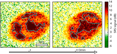
We demonstrated a quantum-enhanced Raman microscope using bright single-beam squeezed light, allowing operation at the optimal Stokes-to-pump ratio for the SRS process. While increased brightness introduced multimode effects that degraded the squeezing, we partially mitigated this using spatial filtering. We applied the microscope to live cell imaging, achieving multispectral, sub-shot-noise images with sufficient speed to observe organelle motion. Quantum correlations provided a modest (~20%) improvement in imaging speed over the shot-noise limit.
Read more:
A. Terrasson, et. al., Fast biological imaging with quantum-enhanced Raman microscopy, Opt. Express, 32, 36193-36206, (2024).